

Growth Plate Injuries in Young Athletes: What Parents Should Know
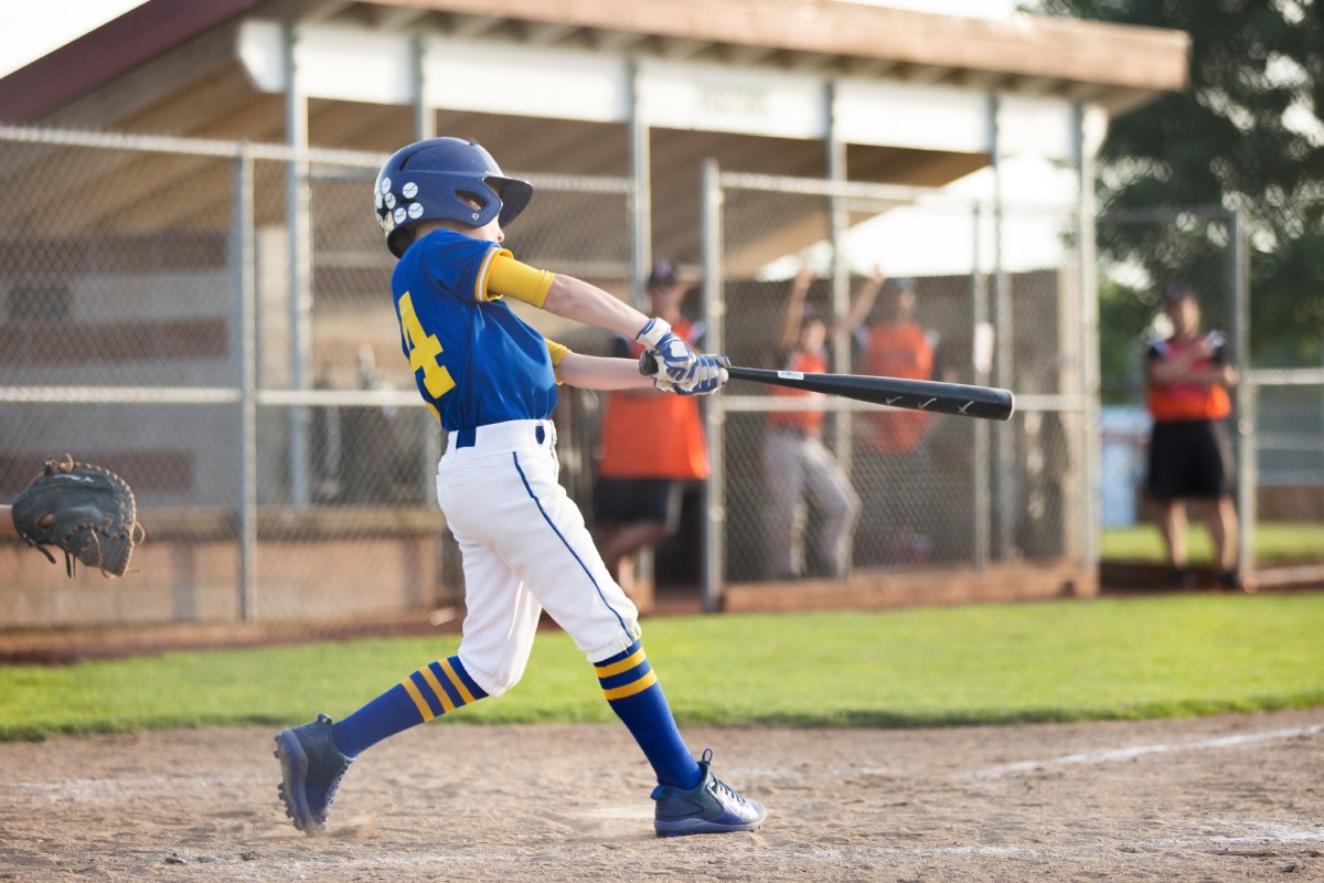
Growth plate injuries are fractures and stress injuries that occur exclusively in children and adolescents, affecting the soft cartilage zones near the ends of long bones where new bone tissue is still forming. Because the physis (the growth plate) is structurally weaker than the surrounding ligaments and tendons during development, the same force that produces a sprain in an adult often causes a fracture in a child.
Knowing the difference matters. Growth plate fractures that are left untreated, misread as soft tissue injuries, or returned to sport too soon can interfere with normal bone growth, with consequences that outlast childhood. However, when these injuries are caught early and managed correctly, most young athletes recover fully and get back to the sports they love.
This piece covers what growth plates are, how to recognize a possible injury, what the treatment process looks like, and when it's time to see an orthopedic specialist rather than waiting to see how the weekend goes.
What Is a Growth Plate and Why Is It Vulnerable?
A growth plate, also called the physis or epiphyseal plate, is a layer of developing cartilage located near the ends of long bones in children and adolescents. It’s where bone elongation happens during childhood. Once a child reaches skeletal maturity, typically in the mid-to-late teenage years, the growth plates close and are replaced by solid bone. Until that happens, these are the softest and most vulnerable parts of a child's skeleton.
The growth plate complex includes three anatomical zones that work together: the epiphysis (the rounded end of the bone), the physis (the cartilage growth layer itself), and the metaphysis (the widening shaft just below the growth plate). Each zone plays a different role in normal bone growth and responds differently to applied force.
Growth plates are present in nearly every long bone in the body: the femur, tibia, radius, and even the small bones of the fingers and toes. In young athletes, the locations that matter most clinically are the distal femur (the frequent site of knee growth plate injuries), the proximal tibia, the proximal humerus (shoulder), and the distal radius (wrist). These are the sites that take the most load in the sports kids play. Early single-sport specialization and year-round training have significantly increased overuse-related growth plate stress, according to the American Academy of Orthopaedic Surgeons, and it's a pattern we see at Alexander Orthopaedics in young athletes across the Tampa Bay area.
What Causes Growth Plate Injuries During Sports?
Acute Traumatic Fractures
Acute growth plate fractures happen when a sudden force—think a collision in football, a hard fall in gymnastics, or an awkward landing—exceeds what the developing physis can absorb. Because growth plate cartilage is less resistant to shear and torsional forces than the surrounding bone and ligament tissue, it gives way first. This is why your child's ankle injury from rolling off a curb or pivoting on the soccer field may turn out to be a growth plate fracture rather than a sprain, even when it looks and sounds identical to what would produce a sprain in an adult.
Contact and collision sports carry the highest risk of acute fracture. Football, wrestling, soccer, and basketball account for the majority of pediatric fracture-related emergency department visits, with football producing injury rates of 4.36 per 1,000 athlete-exposures—the highest of any tracked sport.
Overuse Injuries From Repetitive Stress
Not all growth plate injuries come from a single moment of impact. Growth plate injuries from overuse develop gradually when repetitive mechanical stress on the physis accumulates faster than the tissue can repair itself. The result is inflammation, microfracturing, or progressive widening of the growth plate. It’s a pattern frequently seen in throwing sports (sometimes called “Little League shoulder” and “Little League elbow”), gymnastics (distal radius stress injuries from bearing weight on extended wrists), and distance running.
These injuries are especially easy to miss. Children under 10 are most likely to sustain growth plate stress injuries, and overuse presentations in youth athletes are significantly underreported because the pain often doesn't force the child off the field immediately. What parents often describe as persistent soreness that comes and goes is sometimes a developing physeal stress injury that warrants a closer look.
The Salter-Harris Classification: What Type of Fracture Is It?
When an orthopedic surgeon evaluates a growth plate fracture, one of the first things they determine is where the fracture occurred within the growth plate complex. That's what the Salter-Harris classification system comes in. First proposed in 1963 by Robert Salter and W. Robert Harris, it remains the accepted standard in North American orthopedics. The system sorts fractures into five types based on which anatomical zones are involved and how much the physis itself is at risk.
As a parent, you don't need to memorize this, but understanding the basics helps explain why your child's treatment plan looks the way it does.
- Type I: The fracture runs through the physis only, with no bone fragment involved. It is often invisible on X-ray. The diagnosis rests on point tenderness directly over the growth plate during a physical exam. Swelling and an inability to bear weight are the key signs.
- Type II: The most common type. The fracture passes through the physis and into the metaphysis, producing a triangular bone fragment visible on imaging. With appropriate immobilization, the prognosis is generally excellent.
- Type III: The fracture extends through the physis and into the epiphysis, crossing the joint surface. More precise management is required to restore joint congruity and protect bone growth, which is why imaging and specialist involvement are necessary.
- Type IV: The fracture crosses all three zones: metaphysis, physis, and epiphysis. This carries a higher risk of growth disturbance and frequently requires surgery.
- Type V: A crush injury of the physis from axial compression. Often not visible on initial X-ray and carries the highest risk of premature growth plate closure, limb length discrepancy, and angular deformity.
The general rule is that higher Salter-Harris types carry greater risk to normal bone growth. Physeal complications, including growth arrest and premature closure, occur in 2% to 14% of growth plate injuries overall, with rates climbing significantly at specific locations. Distal tibia fractures result in premature physeal closure in 27.2% of cases. Even lower-type fractures that are poorly managed or caught late can produce growth problems that persist into adulthood. That's why early action matters.
Key Signs and Symptoms Parents Shouldn’t Dismiss
Growth plate injury symptoms in a young athlete include localized pain and tenderness directly over a joint or near the end of a long bone, visible swelling, warmth, and reluctance or inability to bear weight on the affected limb. The early overuse pattern is easy to overlook: pain often appears after activity and resolves with rest, then gradually starts showing up again during further activity.
One of the most common mistakes parents make is treating a growth plate injury as a sprain. The child may not remember a specific moment of injury, particularly with overuse presentations. Any persistent joint pain in a growing child that doesn’t resolve within a few days of rest should be evaluated with imaging, not managed at home indefinitely.
The following symptoms require same-day evaluation and shouldn’t wait:
- Inability to bear weight after an acute injury
- Visible deformity of the limb
- Swelling that develops rapidly in the first hour after injury
- Pain severe enough to prevent sleep
If your child is showing any of these signs after a game, practice, or fall, call your orthopedic provider before the next training session.
How Growth Plate Injuries Are Diagnosed
Clinicians diagnose growth plate fractures through a combination of physical exam, patient history, and imaging tests. X-rays are the standard first-line imaging study and will clearly identify most Type II, III, and IV fractures. Type I fractures, which involve only the physis without a bone fragment, can appear completely normal on X-ray. In those cases, the diagnosis comes from the physical exam: point tenderness directly over the growth plate is the key finding.
MRI (magnetic resonance imaging) is the most sensitive imaging tool for growth plate injuries. It’s used when X-rays are negative or inconclusive, when a Type V crush injury is suspected, or when the full extent of cartilage damage needs to be understood before treatment is planned. CT scan (computed tomography) provides detailed cross-sectional bone imaging and is particularly useful for evaluating complex, displaced, or intra-articular fractures before surgery.
If your child's pediatrician has assessed a joint injury and cleared it as a sprain, but the pain persists beyond a few days without improvement, ask for an orthopedic referral. The longer a displaced or partially displaced fracture goes unrecognized, the greater the risk of malunion and growth disturbance.
Treatment: From Cast to Surgery
Immobilization and Conservative Treatment
Most Type I and Type II growth plate fractures are treated conservatively with immobilization in a cast or splint, rest, and protected weight-bearing. The goal is to maintain anatomical alignment while the physis heals. A well-aligned, non-displaced fracture in a healthy child typically heals within three to six weeks, followed by close imaging to confirm alignment is maintained and bone growth is proceeding normally.
For overuse injuries, the treatment conversation often starts with an uncomfortable truth: a meaningful reduction in training load, not a few days off. Returning to sport before a physeal stress injury has fully recovered is one of the most common reasons these injuries progress from a manageable stress response to a fracture that requires immobilization.
Surgical Intervention for Displaced Fractures
Type III, IV, and some displaced Type II fractures require reduction to restore anatomy. Closed reduction realigns the fracture without opening the skin. It’s typically performed under anesthesia, then the fracture is stabilized with a cast or splint. When closed reduction cannot achieve or hold adequate alignment, open reduction is required: a surgical procedure in which the fracture is exposed directly, reduced under visualization, and held in place with hardware such as wires, screws, or pins.
The primary goal of surgery is restoring anatomical alignment while minimizing additional disruption to the physis. Wires and smooth pins that cross the growth plate are preferred over threaded hardware where possible, as they cause less damage to the cartilage growth layer.
Recovery Timeline: What Parents Should Expect
Recovery from a growth plate fracture follows three distinct phases: initial healing and immobilization, rehabilitation and strength rebuilding, and gradual return to sport. How long each phase takes depends on the fracture type, location, severity, and your child's age and skeletal maturity at the time of injury.
Even after the cast comes off, a physical therapist should guide the return-to-sport progression. Bone healing and functional recovery are not the same thing. Rebuilding strength, range of motion, and neuromuscular control in the affected limb takes additional weeks beyond the bone healing window. Clearance from the treating orthopedic surgeon, not the absence of pain alone, is the right standard for returning to competitive play.
Growth monitoring is part of the long-term plan for any significant growth plate injury. For Type III through V fractures, or any injury where the physis was meaningfully disrupted, follow-up imaging at regular intervals is how the treating physician confirms that bone growth is proceeding normally. Research on distal femoral physeal fractures found premature physeal closure in 23% of cases, which is exactly why this follow-up matters long after your child feels fine.
When to Consult an Orthopedic Specialist
If your child has persistent joint pain after a sports injury—pain that is not improving with rest after a few days, or that keeps coming back with activity—an orthopedic evaluation is the right next step. You don’t need to wait until the pain is unbearable or until something is visibly wrong.
Growth plate fractures and injuries require evaluation and management by an orthopedic surgeon, not observation at home alone for anything beyond the mildest presentations. Delayed diagnosis is a documented driver of growth disturbance and malunion, with potential consequences including limb length discrepancy, angular deformity, and premature joint arthritis in a child who still has years of growing ahead of them.
At Alexander Orthopaedics, we provide pediatric orthopedic and sports medicine care across the Tampa Bay area, with orthopedic surgeons who understand the specific demands of young athletes and the biology of the developing skeleton. If your child has persistent joint pain after a sports injury, visible swelling that does not resolve, or a limp that continues beyond 48 to 72 hours, schedule an evaluation before the next practice or game. Early answers lead to better outcomes.
FAQs
How Serious Is a Growth Plate Fracture?
The seriousness of a growth plate fracture depends on its Salter-Harris type and location. Type I and II fractures (the most common) typically heal well with immobilization and carry a low risk of growth disturbance when treated promptly. Type III, IV, and V fractures involve the joint surface or compress the physis, raising the risk of premature growth plate closure, limb length discrepancy, or angular deformity. Early, accurate diagnosis significantly changes the outcome.
Can Adults Suffer From Growth Plate Fractures?
No. Growth plate fractures occur exclusively in growing children and adolescents because the physis is present only in skeletally immature individuals. Once growth plates close, typically in girls by age 14 to 16 and in boys by age 16 to 18, the cartilage is replaced by solid bone and the vulnerability disappears. An adult sustaining the same force as a child would typically suffer a ligament sprain rather than a physeal fracture.
Can a Growth Plate Fracture Cause Long-Term Problems?
It can, particularly when diagnosis is delayed or treatment is inadequate. Potential long-term complications include growth arrest, limb length discrepancy, angular bone deformity, and early-onset joint arthritis in the affected limb. Physeal complications occur in 2% to 14% of patients after growth plate injury overall, with higher rates at specific locations such as the distal tibia and distal femur. Close follow-up with an orthopedic surgeon through the remainder of your child's growth period is appropriate after any significant growth plate injury.